TAU researchers develop an AI eye scan technique to detect anemia and assess key blood markers
Technique is a step toward needle-free blood testing
Support this researchResearchers from Tel Aviv University (TAU) and Sheba Medical Center have developed an artificial intelligence-based system that can detect anemia and estimate key blood markers using a brief video scan of blood vessels in the eye, potentially eliminating the need for traditional blood draws.
In a proof-of-concept study involving 224 participants, the technology achieved 82.8% accuracy in detecting anemia by analyzing blood-flow patterns in the eye’s conjunctiva. The research represents a promising step toward faster, more accessible, and non-invasive health screening that could one day be used in clinics, community settings, and even at home.
The study was conducted by Tamir Denis, a master’s graduate of TAU, in collaboration with the research groups of Professor Haim Suchowski of the School of Physics and Astronomy and Professor Lior Wolf of the Blavatnik School of Computer Science and AI, together with researchers from Sheba Medical Center. The findings were published on April 8, 2026, in the scientific journal npj | Digital Medicine.
Blood tests are among the most commonly performed medical procedures worldwide, yet they still rely on invasive blood sampling and complex laboratory processing. Previous attempts focused on anatomical sites failed to demonstrate significant correlation. The researchers note that this new technology could eventually enable faster and more accessible testing, particularly in regions where access to healthcare services is limited. The study highlights that anemia is one of the most prevalent medical conditions in the world, affecting approximately 30% of the global population.
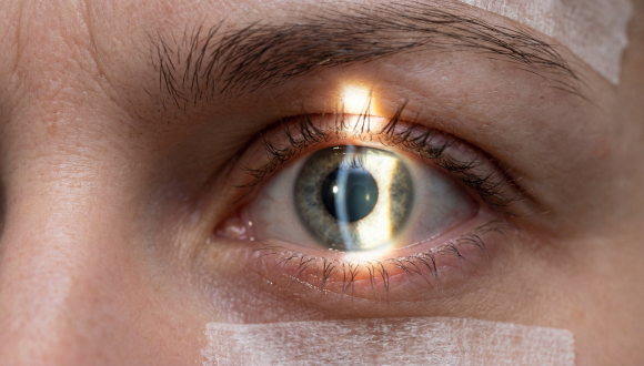
The study presents a technology called Video-to-Vessels, which converts high-magnification video recordings of the tiny blood vessels in the eye’s conjunctiva into a compact digital representation of vascular structure and blood-flow dynamics. This information is then fed into an artificial intelligence system trained to identify correlations between blood-flow characteristics and key blood markers, such as hemoglobin (Hb) levels and red blood cell (RBC) counts.
The study included 224 participants who underwent both standard blood tests and imaging of the conjunctiva using a 50x magnification RGB camera. Ten-second video recordings were collected from both eyes of each participant.
The study’s principal finding was that the system achieved a relatively high accuracy rate of 82.8% in detecting anemia. In addition, a strong correlation was found between the system’s predictions and laboratory results for both hemoglobin levels and red blood cell counts.
According to the researchers, one of the most notable examples of the system’s capabilities was its ability to detect subtle differences among extremely thin blood vessels. The study found that these vessels provided the most accurate information for predicting hemoglobin levels. The researchers explain that in very narrow vessels, blood cells move in single file, making it easier to identify blood-flow patterns and changes associated with hemoglobin concentration. Models trained exclusively on these small vessels achieved significantly better results than those based on larger vessels.
Another key finding was the importance of video processing. The researchers demonstrated that stabilizing eye movements and removing digital noise significantly improved the system’s performance. When these steps were omitted from the process, the predictive correlation declined by 38% for hemoglobin and by 19% for red blood cell counts.
The researchers emphasize that this remains a proof-of-concept study, and that broader, more diverse studies will be needed before the technology can be implemented in clinical practice. Nevertheless, they believe that it could eventually be developed into a compact handheld device to serve as a first-line screening tool in clinics, community healthcare settings, and even at home.